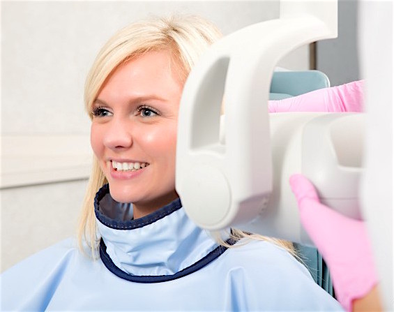
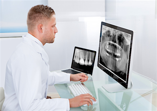
Nowadays, dentistry cannot do without diagnostic radiology also known as X-rays. The images of intraoral basic radiography, in 3-D or panoramic will give your dentist valuable information on your teeth and gums, and help him choose the best treatment for dental patients.
Many worry that dental X-ray equipment emits harmful radiation for humans. In fact, measurements of radiation transmitted during the dental radiology process show that they are no different from those to which we are exposed daily, such as boarding an aircraft, and the proximity to televisions and smoke detectors.
Myth # 1
“Do I need an X-ray every time I visit the dentist? ”
Reality
The frequency with which you may need X-rays will depend on your oral health. A healthy adult who hasn’t had cavities or other problems for a few years does not probably need X-rays at every appointment.
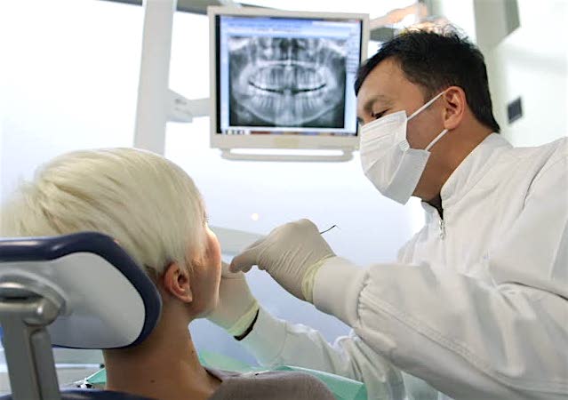
For most people, an examination every 6 months is enough, but the frequency of your visits and X-rays will depend on your dental needs and your dentist will guide you according to your personal hygiene.
The purpose of an examination is to identify hidden problems early. X-rays are the only way to see the inside of tissues and detect things undetectable to the naked eye.
Myth # 2
“Too many dental X-rays can cause brain tumors.”
Reality
A few years ago, the Order of Quebec Dentists refuted the findings of a 2012 study on which is based the article “Dental X-Rays and Risk of Meningioma”. According to the ODQ, the article does not prove a causal relationship between ionizing radiation of dental origin and the development of meningioma.
This article by Yale University epidemiologists found that dental X-rays increase the risk of meningioma, a benign brain tumor. For example, the researchers argue that dental X-rays are the main artificial source of exposure to ionizing radiation in the United States, a form of radiation associated with an increased risk of brain tumors.
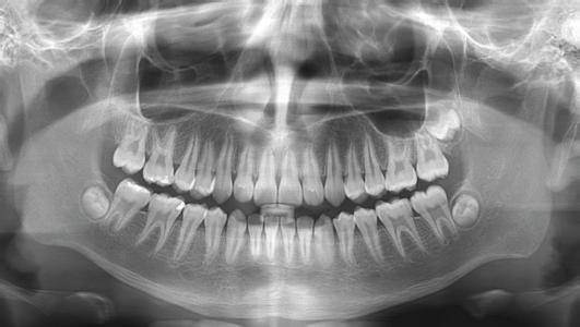
Less risk with 3D radiography
3D digital radiography equipment limit patient exposure time with X-rays, while ensuring higher precision images of the focus area. Because of this, anatomical depth and relationships between different dental structures are identified and measured accurately for more reliable diagnostics and better treatments.

Myth # 3
“Never expose a pregnant woman or a child to radiography.”
Reality
There is no medical indication against taking dental X-rays on pregnant patients. However, at the request of the patient, elective procedures may be postponed until after pregnancy.
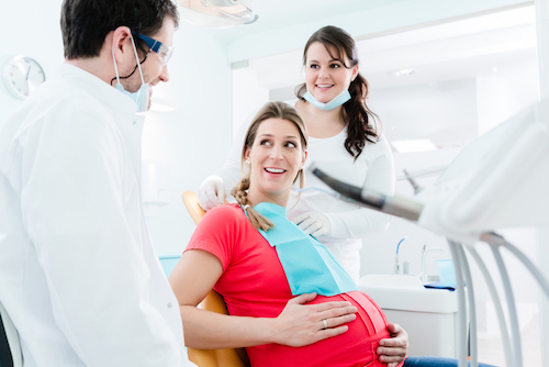
If you are pregnant and you must have an X-ray, the dental assistant will have you wear a lead apron to the front and a lead collar for the thyroid to protect vulnerable body parts.
X-rays for children?
Radiography will provide information about your child’s condition that isn’t visible during a clinical oral evaluation.
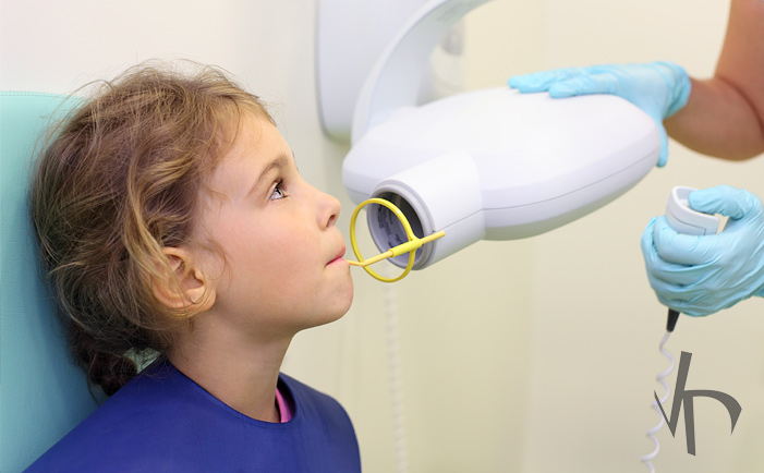
It’s impossible to sign a waiver exempting X-rays for your child if they are necessary to conclude a diagnosis for an orthodontic problem and to plan the best treatment needed.
Myth # 4
“Dental personnel risk exposure to ionizing radiation.”
Reality
Even if we are aware of all the benefits of patients X-raying, care must be taken to reduce the potentially harmful effects of ionizing radiation for the health of X-ray operators.

Reducing risk for staff
Dental personnel can reduce the risks associated with radiation exposure by following established security measures and by carrying a dosimeter. This is a device that measures the degree of X-ray exposure during a given period. An exposure report indicating the radiation dose received during this period is then produced.
At the Clinic, we maintain a log of the number of radiographs taken daily and we submit this information to the Public Health Agency of Canada laboratory when renewing our dental radiology permit. We must also send dosimeters periodically to the national dosimetry service.
Continuous training of operators
Our staff has completed the training necessary to operate our radiographic equipment. Manufacturers provide on-site training during installation, and they come back to update the training required by new equipment technology.
Myth # 5
“X-rays on film were more reliable than digital counterpart today.”
Reality
David Côté Dental Clinic replaced film X-rays by digital radiography over 15 years ago. This technique offers unmatched precision and allows even sharper diagnoses. Unlike the old method, the digital images are stored on the clinic’s computer system.
In addition, the radiation level with digital is significantly lower than the traditional method. This technique also allows instant results and rapid email transmission to relevant bodies. A plus, having eliminated the use of plastic film is very beneficial to the environment.
Digital benefits
- Digital radiography will keep images in your electronic dental record.
- 90% reduction of radiation exposure levels to traditional X-rays.
- Instant results mean less waiting for our patients.
- Quick emailing of digital X-rays to insurance companies and dentists.
- Protects the environment by eliminating toxic solutions for film development.
Conclusion
At our Clinic, we are committed to making our patients’ dental experience as safe and as comfortable as possible. We keep the radiation dose to which a patient is exposed to a minimum, while ensuring adequate radiological examinations and diagnoses.
We hope this article has shed light on a subject that can cause anxiety in some patients. Once again, we thank you for reading and sharing our blog articles with friends! Floating Share Buttons are on the left of articles.
You have more questions about the use of dental X-rays? Ask Dr. Dubois or Dr. Côté, they will be more than happy to answer you.

